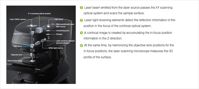
This technique captures the surface image and enables magnification control by reducing the scan area to smaller sizes thus increasing magnification without any resolution loss.
Laser scanning microscope magnification.
Light microscope transmission electron microscope scanning electron microscope and laser scanning confocal microscope.
Clsm combines high resolution optical imaging with depth selectivity which allows us to do optical sectioning.
A the use of microscopy to observe and investigate different types of cell and cell structure in a range of eukaryotic organisms to include an appreciation of the images produced by a range of microscopes.
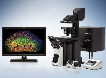
With confocal laser scanning microscopy clsm we can find out even more.
Fred brakenhoff developed a scanning confocal microscope in 1979 21 while almost simultaneously colin sheppard contributed to the technique with a theory of image formation 22.
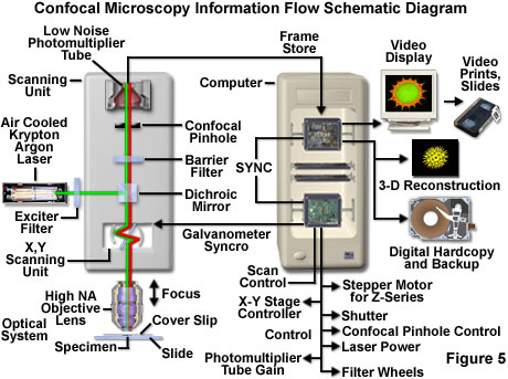
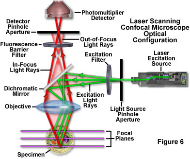
Confocal microscopy most frequently confocal laser scanning microscopy clsm or laser confocal scanning microscopy lcsm is an optical imaging technique for increasing optical resolution and contrast of a micrograph by means of using a spatial pinhole to block out of focus light in image formation.
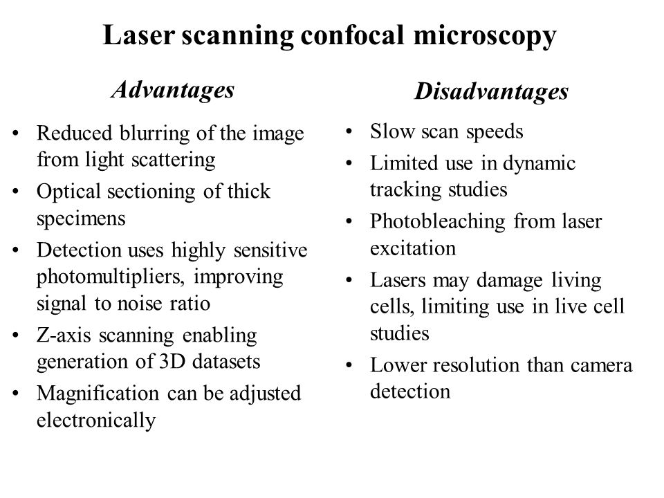
The confocal laser scanning microscope s aim was not to further increase magnification but to make clearer.
Laser scanning confocal microscopy laser scanning confocal microscopes employ a pair of pinhole apertures to limit the specimen focal plane to a confined volume approximately a micron in size.
Capturing multiple two dimensional images at different depths in a sample enables the.
In the past the traditional laser microscope excited the whole thickness of the sample resulting in saturated blurry images and sometimes visualizing false colocalization images.
The laser scanning microscope uses a scanning design called beam scanning where the laser image path is scanned in a raster pattern on the surface of the sample.
Relatively thick specimens can be imaged in successive volumes by acquiring a series of sections along the optical z axis of the microscope.
The confocal laser scanning microscope clsm is a microscope which focuses only on a single focal plane and the unfocused plane will not be visualized.
Fluorescent microscopy not only makes our images look good it also allows us to gain a better understanding of cells structures and tissue.
.jpg?rev=33A9)


