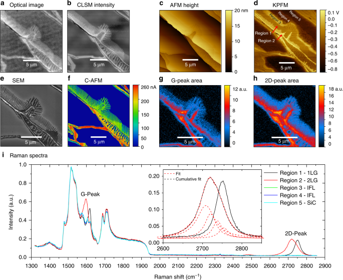
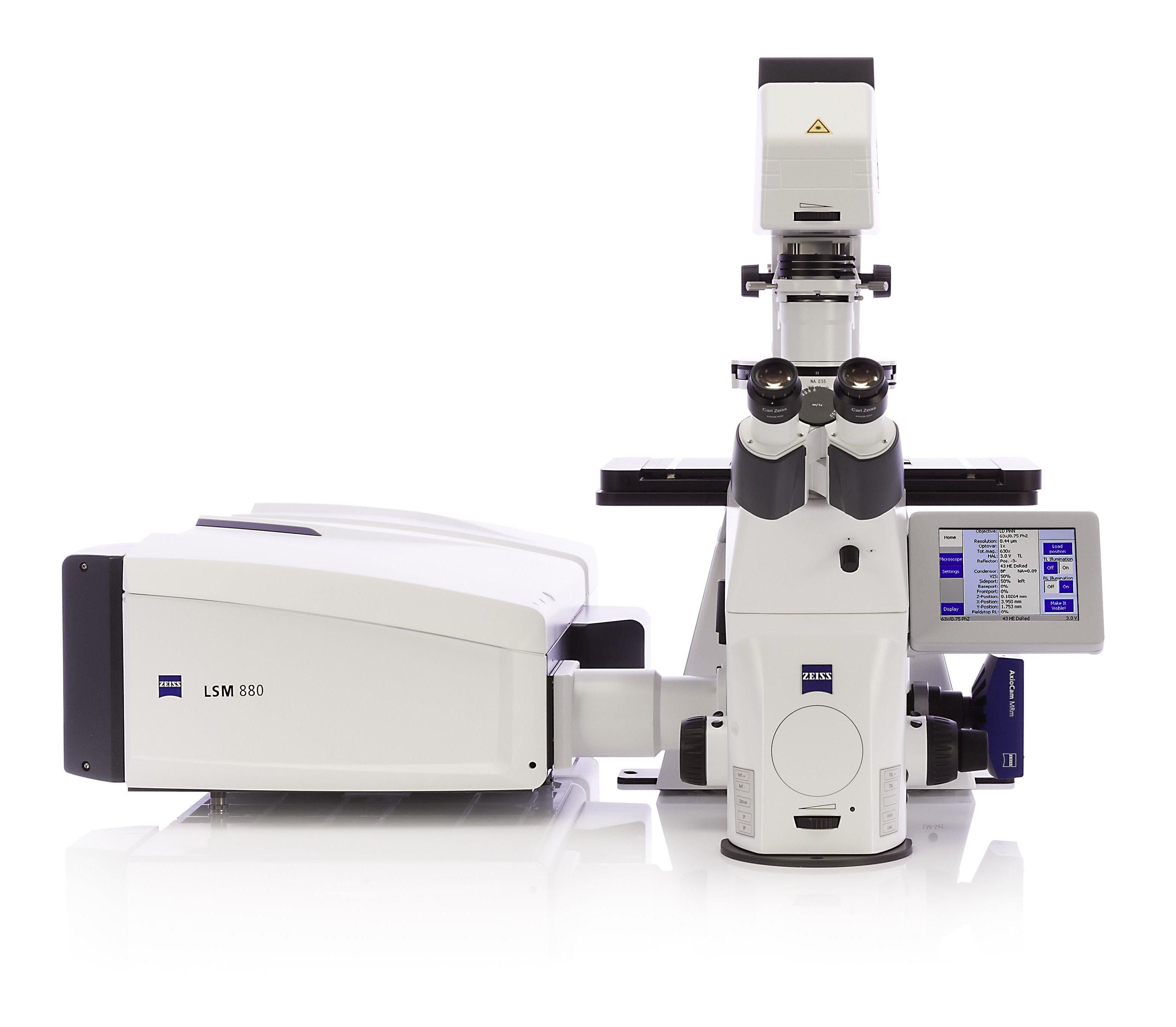
In the past the traditional laser microscope excited the whole thickness of the sample resulting in saturated blurry images and sometimes visualizing false colocalization images.
Laser scanning microscope maximum magnification.
Capturing multiple two dimensional images at different depths in a sample enables the.
This feature is termed the zoom factor and is usually employed to adjust the image spatial resolution by altering the scanning laser sampling period.
Lets you look specifically at parts of a cell such as individual proteins by labelling them with fluorescence.
With confocal laser scanning microscopy clsm we can find out even more.
Clsm combines high resolution optical imaging with depth selectivity which allows us to do optical sectioning.
Lens used in a lm.
A confocal laser scanning microscope or also known as laser scanning confocal microscope is used to obtain high resolution images and generate 3d reconstructions through direct optical sectioning to provide clear images from a range of depths of thicker specimens for nanometer level imaging and measurement.
The confocal laser scanning microscope clsm is a microscope which focuses only on a single focal plane and the unfocused plane will not be visualized.
Relatively thick specimens can be imaged in successive volumes by acquiring a series of sections along the optical z axis of the microscope.
Lscm laser scanning confocal microscope parameter value comment maximum ip pixel resolution 1270 x 1000 pixel user adjustable maximum field of view 1 ccd equivalent 9 6 x 12 8mm.
The maximum magnification of a light microscope.
Fluorescent microscopy not only makes our images look good it also allows us to gain a better understanding of cells structures and tissue.
It has a maximum magnification of about 100000.
This means that we can view visual sections of tiny structures that.
The maximum magnification of a tem.
Confocal microscopy most frequently confocal laser scanning microscopy clsm or laser confocal scanning microscopy lcsm is an optical imaging technique for increasing optical resolution and contrast of a micrograph by means of using a spatial pinhole to block out of focus light in image formation.
Confocal laser scanning fluorescence microscope.
Light microscope laser scanning confocal microscope transmission electron microscope tem and scanning electron microscope sem light.
Laser scanning confocal microscopy laser scanning confocal microscopes employ a pair of pinhole apertures to limit the specimen focal plane to a confined volume approximately a micron in size.
A transmission electron microscope tem produces a 2d image of a thin sample and has a maximum resolution of 500000.
A scanning electron microscope sem produces a 3d image of a sample by bouncing electons off and dectecting them at multiple detectors.


